Scanning Electron Microscopy Lab
“Automated Mineralogy Lab”
Building T – Room T 030
Tel: +49 30 838 70823
Contact: Dr. J. C. Vrijmoed
Email: j.c.vrijmoed@fu-berlin.de
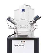
The Zeiss Sigma 300 VP Field-Emission Scanning Electron Microscope is equipped with:
- Zeiss Gemini column
- 2 Bruker Quantax Xflash 60mm2 SDD EDS Detectors for quantitative Element Analysis
-
1 Variable Pressure Secondary Electron detector (VPSE)
-
1 High Definition Back Scatter Detector (HDBSE)
-
1 Inlens Detector
-
2 Zeiss ATLAS Correlative microscopy system (1 online/1 offline Workstation)
-
2 Mineralogic Mining automated Mineralogy systems (1 online/1 offline Workstation)
-
2 Reservoir Mining automated Pore analysis systems (1 online/1 offline Workstation)
Applications
-
High-resolution automated surface imaging (Zeiss ATLAS)
-
Surface morphology imaging of uncoated, unpolished samples (Low Vac)
-
Automated quantitative Mineralogy- and Porosity mapping of thinsections
-
Fast automated search of specific Mineral Phases (i.e. Sulfides, Zircons)
-
Grain size/shape analysis and Mineral paragenesis analysis
-
Cathodoluminescence analysis (with VPSE Detector)
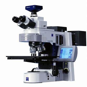
The ZEISS Axio Imager M2m is optimized for automated transmitted/reflected light thinsection mapping. It is equipped with:
-
4 Objectives 2.5x, 5x, 10x, and 20x (optimized for reflected light)
-
Polarized transmitted light / motorized reflectors for polarized reflected light
-
Motorized stage
-
High Definition digital camera
-
Zen Blue Software
-
High performance PC for the analysis of GB-sized image data
Applications
-
Fast full thinsection mapping for the correlation with SEM images and EDS data
-
Fast thin section search for opaque phases like Sulfides, Oxides, etc.